lv pseudoaneurysm vs true aneurysm radiology | lv aneurysm post mi lv pseudoaneurysm vs true aneurysm radiology The diagnosis of left ventricular pseudoaneurysm is a challenge. Imaging features that help to differentiate false from true aneurysms include the neck-to-body diameter ratio . Dos. Clean with a damp cloth and slightly soapy water to wipe down the canvas. Micellar water and baby wipes are gentle cleaning alternatives you can use as well. Condition your bag every 6 – 12 months (or depending on how often you use your LV canvas bag). To prevent cracking, we recommend using apple conditioner cream and .
0 · true aneurysm vs false
1 · pseudoaneurysm vs true aneurysm echo
2 · lv aneurysm vs pseudoaneurysm echo
3 · lv aneurysm post mi
4 · lv aneurysm on echo
5 · left ventricular pseudoaneurysm vs aneurysm
6 · left ventricular aneurysm post mi
7 · left ventricular aneurysm guidelines
This projector is a WXGA model capable of providing light output of 3,000 lumens, which is bright for a projector in portable class. Its contrast ratio is 2300:1 to project sharp images. Ultra short-throw projector that enables close-in projection onto a screen or wall.$599 (USD) Status. Discontinued Jul 2016. Released. October 2014. Warranty. 3 Years. User Reviews. Review this Projector. Switch to Metric. Brightness. 3,000 Lumens (ANSI) 1 / 2,100 Lumens (Eco) Resolution. 1280x800. Aspect Ratio. 16:10 (WXGA) Contrast. 2,300:1 (full on/off) Display Type. 0.65" DLP x 1. Color Processing. 8-bit.
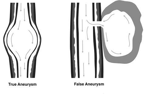
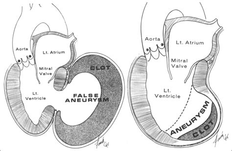
A true aneurysmal sac contains an endocardium, epicardium, and thinned fibrous tissue (scar) which is a remnant of the left ventricular muscle, while a pseudoaneurysm sac represents a pericardium that contains a ruptured left ventricle 5.
LV pseudoaneurysm is formed if cardiac rupture is contained by pericardium, organizing thrombus, and hematoma. This condition calls for urgent surgical repair. Whereas, in a true .
true aneurysm vs false
pseudoaneurysm vs true aneurysm echo
MATERIALS AND METHODS: Cardiac MR images obtained in 22 sequential patients (20 men, two women; mean age, 63 years; age range, 45–75 years) with pathologically proved left . Differentiation between LV pseudoaneurysms and true aneurysms can be challenging and investigations include transthoracic echocardiography/transoesophageal . The diagnosis of left ventricular pseudoaneurysm is a challenge. Imaging features that help to differentiate false from true aneurysms include the neck-to-body diameter ratio .The tissue characterization of cardiac MRI make it ideal for evaluation of pseudoaneurysm of the ventricles and for distinguishing pseudoaneurysm from true aneurysms. The use of late .
Left ventricular (LV) pseudoaneurysms form when cardiac rupture is contained by adherent pericardium or scar tissue (1). Thus, unlike a true LV aneurysm, a LV pseudoaneurysm .A postmyocardial infarction left ventricular pseudoaneurysm occurs when a rupture of the ventricular free wall is contained by overlying, adherent pericardium. A postinfarction . Left ventricular (LV) aneurysms and pseudoaneurysms are two complications of myocardial infarction (MI) that can lead to death or significant morbidity. This topic reviews the . Most of the LVPs develop after MI or cardiothoracic surgery. In a systematic literature review of 290 patients, MI (55%), surgery (33%), and trauma (7%) were the top 3 .
A true aneurysmal sac contains an endocardium, epicardium, and thinned fibrous tissue (scar) which is a remnant of the left ventricular muscle, while a pseudoaneurysm sac represents a pericardium that contains a ruptured left ventricle 5.LV pseudoaneurysm is formed if cardiac rupture is contained by pericardium, organizing thrombus, and hematoma. This condition calls for urgent surgical repair. Whereas, in a true aneurysm, LV out-pouching is a thinned out wall but with some degree of myocardium wall integrity intact. Such an entity calls for elective surgery.MATERIALS AND METHODS: Cardiac MR images obtained in 22 sequential patients (20 men, two women; mean age, 63 years; age range, 45–75 years) with pathologically proved left ventricular true aneurysm (n = 18) or false aneurysm (n = 4) after myocardial infarction were retrospectively analyzed.
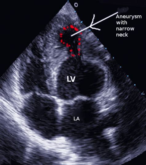
Differentiation between LV pseudoaneurysms and true aneurysms can be challenging and investigations include transthoracic echocardiography/transoesophageal echocardiography, LV angiography, magnetic resonance imaging, computed tomography, radionuclide scanning. The diagnosis of left ventricular pseudoaneurysm is a challenge. Imaging features that help to differentiate false from true aneurysms include the neck-to-body diameter ratio (smaller in false aneurysm); 5 distribution of the aneurysmal sac; and discontinuity of myocardium at the neck of the aneurysm.The tissue characterization of cardiac MRI make it ideal for evaluation of pseudoaneurysm of the ventricles and for distinguishing pseudoaneurysm from true aneurysms. The use of late gadolinium enhancement to identify the location and transmural extent of prior infarcts is particularly valuable [ 12 ].
Left ventricular (LV) pseudoaneurysms form when cardiac rupture is contained by adherent pericardium or scar tissue (1). Thus, unlike a true LV aneurysm, a LV pseudoaneurysm contains no endocardium or myocardium (2).A postmyocardial infarction left ventricular pseudoaneurysm occurs when a rupture of the ventricular free wall is contained by overlying, adherent pericardium. A postinfarction aneurysm, in contrast, is caused by scar formation resulting in thinning of the myocardium. Left ventricular (LV) aneurysms and pseudoaneurysms are two complications of myocardial infarction (MI) that can lead to death or significant morbidity. This topic reviews the diagnosis and management of patients with aneurysms or pseudoaneurysms caused by MI. Most of the LVPs develop after MI or cardiothoracic surgery. In a systematic literature review of 290 patients, MI (55%), surgery (33%), and trauma (7%) were the top 3 associations. 1 LVPs carry a substantial risk of rupture, which is considerably higher than that of a .
lv aneurysm vs pseudoaneurysm echo
A true aneurysmal sac contains an endocardium, epicardium, and thinned fibrous tissue (scar) which is a remnant of the left ventricular muscle, while a pseudoaneurysm sac represents a pericardium that contains a ruptured left ventricle 5.LV pseudoaneurysm is formed if cardiac rupture is contained by pericardium, organizing thrombus, and hematoma. This condition calls for urgent surgical repair. Whereas, in a true aneurysm, LV out-pouching is a thinned out wall but with some degree of myocardium wall integrity intact. Such an entity calls for elective surgery.MATERIALS AND METHODS: Cardiac MR images obtained in 22 sequential patients (20 men, two women; mean age, 63 years; age range, 45–75 years) with pathologically proved left ventricular true aneurysm (n = 18) or false aneurysm (n = 4) after myocardial infarction were retrospectively analyzed. Differentiation between LV pseudoaneurysms and true aneurysms can be challenging and investigations include transthoracic echocardiography/transoesophageal echocardiography, LV angiography, magnetic resonance imaging, computed tomography, radionuclide scanning.
The diagnosis of left ventricular pseudoaneurysm is a challenge. Imaging features that help to differentiate false from true aneurysms include the neck-to-body diameter ratio (smaller in false aneurysm); 5 distribution of the aneurysmal sac; and discontinuity of myocardium at the neck of the aneurysm.The tissue characterization of cardiac MRI make it ideal for evaluation of pseudoaneurysm of the ventricles and for distinguishing pseudoaneurysm from true aneurysms. The use of late gadolinium enhancement to identify the location and transmural extent of prior infarcts is particularly valuable [ 12 ].
Left ventricular (LV) pseudoaneurysms form when cardiac rupture is contained by adherent pericardium or scar tissue (1). Thus, unlike a true LV aneurysm, a LV pseudoaneurysm contains no endocardium or myocardium (2).
A postmyocardial infarction left ventricular pseudoaneurysm occurs when a rupture of the ventricular free wall is contained by overlying, adherent pericardium. A postinfarction aneurysm, in contrast, is caused by scar formation resulting in thinning of the myocardium. Left ventricular (LV) aneurysms and pseudoaneurysms are two complications of myocardial infarction (MI) that can lead to death or significant morbidity. This topic reviews the diagnosis and management of patients with aneurysms or pseudoaneurysms caused by MI.


lv aneurysm post mi

lv aneurysm on echo
left ventricular pseudoaneurysm vs aneurysm
left ventricular aneurysm post mi
To cancel your car insurance policy, give us a call: 0330 678 5111 If you have a multi car policy, call us and we can remove the car, without you having to cancel the whole policy.
lv pseudoaneurysm vs true aneurysm radiology|lv aneurysm post mi


